Eʼelyaaígíí:PET-image.jpg

Size of this preview: 679 × 600 pixels. Other resolutions: 272 × 240 pixels | 543 × 480 pixels | 869 × 768 pixels | 1,132 × 1,000 pixels.
''full resolution''ígíí yishʼį́ nisin (1,132 × 1,000 pixels - 139 KIIŁTSOHʼÍÍŁKÉ, MIME type: image/jpeg)
Summary
| Description |
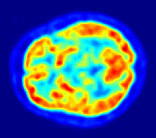
English: This is a transaxial slice of the brain of a 56 year old patient (male) taken with positron emission tomography (PET). The injected dose have been 282 MBq of 18F-FDG and the image was generated from a 20 minutes measurement with an ECAT Exact HR+ PET Scanner. Red areas show more accumulated tracer substance (18F-FDG) and blue areas are regions where low to no tracer have been accumulated.
العربية: صورة مقطعية للدماغ البشري تظهر استهلاك الطاقة. |
||
| Date | |||
| Source | Own work | ||
| Author | Jens Maus (http://jens-maus.de/) | ||
| Permission (Reusing this file) |
|
łahgo ádaalyaaígíí
Ńtʼę́ę́ʼígíí yíníʼį́įgo biniiyé, naʼalkid/yoołkáłígíí bikáaʼgi "click" ádíílííł
| naʼalkid/yoołkáłígíí | thumbnailígíí | naaniigo/náásee | Choyoołʼįįhí | haneʼ | |
|---|---|---|---|---|---|
| kʼadígíí | 02:00, 12 Níłchʼitsoh 2017 | | 1,132 × 1,000 (139 KIIŁTSOHʼÍÍŁKÉ) | SteinsplitterBot | Bot: Image rotated by 270° |
| 14:36, 16 Wóózhchʼį́į́d 2010 |  | 1,002 × 1,132 (134 KIIŁTSOHʼÍÍŁKÉ) | Damato | uploaded another PET image with a higher resolution which might be more usable for printing it and which has a better color scale. | |
| 09:47, 7 Níłchʼitsʼósí 2005 |  | 373 × 405 (48 KIIŁTSOHʼÍÍŁKÉ) | Damato | This is an image taken from a typical PET acquisition. It is a tomographic view of a brain examination in transaxial view. Red areas show more accumulated radioactivity and blue areas are partions where low to no activity was accumulated. It should illust |
''Wikiibíídiiya'' bikáaʼgi choolʼį́/yitʼį́
ałʼąą dineʼé bizaadjí
- ar.wikipedia.org bikáaʼgi
- arz.wikipedia.org bikáaʼgi
- ast.wikipedia.org bikáaʼgi
- bg.wikipedia.org bikáaʼgi
- bn.wikipedia.org bikáaʼgi
- ca.wikipedia.org bikáaʼgi
- de.wikipedia.org bikáaʼgi
- de.wikibooks.org bikáaʼgi
- el.wikipedia.org bikáaʼgi
- en.wikipedia.org bikáaʼgi
- Positron emission tomography
- Neurolinguistics
- Human brain
- Scintigraphy
- Timeline of tuberous sclerosis
- User:Portakalsinatra
- Wikipedia:Wikipedia Signpost/2011-03-07/Features and admins
- User talk:Silver seren/Archive 10
- Childhood acquired brain injury
- User:Rkasinadhuni3/practice sandbox
- User:Mcorrin3/Sandbox Practice
- User:LoriJeanMarie/Brain science practice page
- User:Gilyardterence/Pediatric Acquired Brain Injury
- Wikipedia:Wikipedia Signpost/Single/2011-03-07
- Wikipedia:WikiProject Cannabis/Members
- User:Anthonyhcole/Parkinson's disease
- User:Silver seren/Barnstars
- User:Flyer22 Frozen/Human brain
- User:Cglife.bmarcus/WikiProjectCards/WikiProject Cannabis
- en.wikiquote.org bikáaʼgi
- en.wikiversity.org bikáaʼgi
- es.wikipedia.org bikáaʼgi
View more global usage of this file.